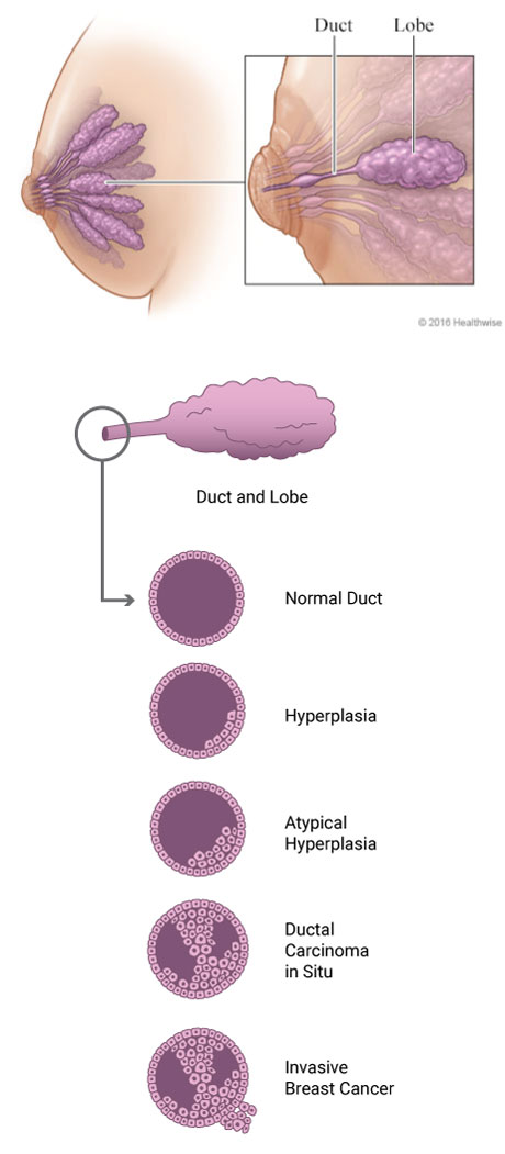
The breast usually has 1 to 2 layers of cells that line the milk ducts and lobules. These cells look the same or similar under a microscope.
In a condition called hyperplasia, the cells grow very quickly and there are too many normal cells. The cells start to build up in the milk ducts and lobules of the breast.
Sometimes these cells begin to change and no longer look the same. When this happens, the cells are called
atypical.
Atypical hyperplasia describes when these cells change and become abnormal. The image below shows how the cells can change over time, becoming more abnormal in size, shape, and appearance.
There are 2 kinds of atypical hyperplasia:
- Atypical ductal hyperplasia is found in the ducts of the breast.
- Atypical lobular hyperplasia is found in the lobules of the breast.
If you have 1 of these conditions, it doesn’t mean you have cancer. It can take many years for atypical cells to become a breast cancer. But you are at higher risk for developing breast cancer.
Breast cancer cells start as non-invasive (in situ). But when they are left undiagnosed or untreated, these cells can invade the surrounding tissues and spread (become invasive or metastatic).
Many people may not know they have atypical hyperplasia. These changes do not show up on a regular screening mammogram, and you can’t feel them during a breast self-exam.
Atypical hyperplasia may be found by your healthcare provider if they are checking a breast lump or nipple discharge or doing a biopsy (tissue sample). If you know you have this condition, talk with your healthcare provider about a treatment and follow-up plan.